
articles

Dry eye is a common condition that occurs when your eyes do not produce enough tears or when the tears evaporate too quickly. This can result in discomfort, irritation, and a gritty sensation in the eyes. Understanding the causes and symptoms of dry eye is crucial for finding effective treatment options. Tyrvaya offers a breakthrough solution for dry eye relief.
What Causes Dry Eye?
There are several factors that can contribute to the development of dry eye. The meibomian glands are responsible for producing the oily component of the tear film, which helps prevent evaporation of tears and maintains a smooth ocular surface. Meibomian gland dysfunction occurs when these glands become blocked, leading to a decrease in the quantity and quality of the meibum. This can result in evaporative dry eye, discomfort, and inflammation of the eyelid margins.
Another common causes is age. As we get older, our tear production tends to decrease, making us more prone to dryness. Hormonal changes, such as those that occur during menopause, can also affect tear production and lead to dry eye.
Environmental factors can play a role as well. Dry or windy climates, air conditioning, and excessive screen time can all contribute to dry eye. Additionally, certain medications, such as antihistamines and antidepressants, can cause dryness as a side effect.
Other underlying health conditions, such as autoimmune diseases like Sjogren's syndrome or rheumatoid arthritis, can also contribute to dry eye. In these cases, the body's immune system mistakenly attacks the tear glands, leading to reduced tear production.

Migraines are a debilitating neurological condition that affect millions of people worldwide. One of the most common and disabling symptoms of migraines is light sensitivity, also known as photophobia. This heightened sensitivity to light can be a significant source of discomfort and pain for those suffering from migraines, making it difficult to function during an attack.
Photophobia is a common symptom of migraines, with up to 80% of migraine sufferers experiencing sensitivity to light during an attack. This sensitivity can be triggered by various types of light, including natural sunlight, fluorescent lighting, and even the glow of computer screens or smartphones.

Clear Lens Extraction (CLE) is a surgical procedure that involves the removal of the natural lens of the eye and replacing it with an artificial lens implant, also known as an intraocular lens (IOL). CLE is primarily performed to correct refractive errors, such as nearsightedness, farsightedness, and astigmatism.
Understanding CLE and How It Works
Clear Lens Extraction is similar to cataract surgery, where the natural lens is removed and replaced with an IOL. However, in CLE, the lens is clear and not clouded as in the case of cataracts. The procedure begins with the administration of local anesthesia to numb the eye and ensure a painless experience for the patient.
Once the eye is numb, a small incision is made on the cornea, which is the clear front surface of the eye. Through this incision, your eye surgeon accesses the lens and carefully removes it. The artificial IOL is then inserted into the empty lens capsule. The IOL is specifically chosen to correct the patient's refractive error, providing them with improved vision.
After the IOL is implanted, the incision is closed with tiny sutures or self-sealing techniques. Following the surgery, patients are usually prescribed antibiotic and anti-inflammatory eye drops to prevent infection and reduce inflammation.
Factors to Consider Before Undergoing Clear Lens Extraction
Before deciding to undergo Clear Lens Extraction, there are several factors that need to be considered. Firstly, it is crucial to have a thorough eye examination to determine if you are a suitable candidate for the procedure. Your optometrist will evaluate your overall eye health and discuss your expectations and goals for vision correction.
Age is another important factor to consider. CLE is typically recommended for individuals over the age of 40 who have developed presbyopia, a condition that affects near vision. It is also important to have stable vision, as any changes in prescription can affect the accuracy of the IOL power calculation.
Additionally, it is essential to understand the potential risks and complications associated with the procedure. While CLE is generally safe, there is a small risk of infection, bleeding, and retinal detachment. Your optometrist will discuss these risks with you and address any concerns you may have.
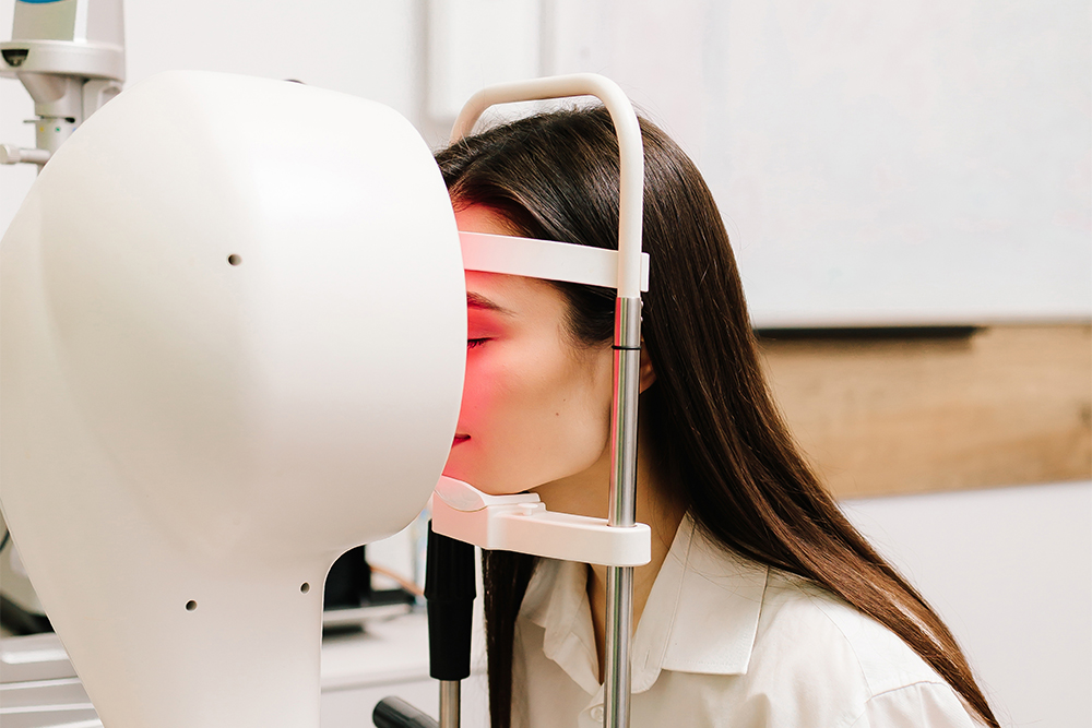
When it comes to diagnosing and managing complex vision conditions, precision matters. One of the most advanced tools is Medmont topography - a state-of-the-art technology that provides highly detailed maps of the cornea. Whether you are being evaluated for specialty contact lenses, dry eye treatment, or refractive surgery, Medmont topography plays a critical role in delivering accurate, customized care.
What Is Medmont Topography?
Medmont topography refers to corneal imaging performed with the Medmont E300 Corneal Topographer, a diagnostic device designed to capture detailed curvature measurements of the cornea.
Using advanced Placido disc technology, the system projects illuminated rings onto the cornea and analyzes their reflections. From this data, it generates a color-coded map that reveals the shape, elevation, and curvature of the cornea with exceptional accuracy.
Why Is Corneal Topography Important?
The cornea is responsible for approximately two-thirds of the eye’s focusing power. Even subtle irregularities can significantly affect vision. Medmont topography allows your eye doctor to detect and monitor:
Keratoconus and other corneal ectasias
Corneal irregularities or scarring
Post-surgical corneal changes
Astigmatism patterns
Contact lens fitting complications
Early signs of corneal distortion
Because it provides highly detailed imaging, Medmont topography often identifies changes long before they become noticeable in a routine vision exam.
How Medmont Topography Improves Contact Lens Fitting
For patients who require specialty contact lenses - such as scleral lenses, hybrid lenses, or lenses for keratoconus - precise corneal mapping is essential.
The Medmont system offers:
High-resolution curvature data
Wide corneal coverage
Repeatable, reliable measurements
Advanced software analysis
This allows your doctor to design and fit custom lenses with greater accuracy, improving comfort, stability, and visual clarity while reducing trial-and-error fittings.

If you find it difficult to tell colors apart, you may be color blind. Color blindness, or color deficiency, is estimated to affect around 8% of men and about 1% of women, but for those affected, it can significantly impact the quality of their day-to-day life. Contrary to popular belief, being color blind doesn’t mean that you can’t see any color at all. Instead, patients simply struggle to differentiate between certain colors. The vast majority of people who are color blind find it impossible to tell the difference between varying shades of red and green. You may hear this referred to as red-green color deficiency. However, this doesn’t only mean that they mix up red and green. They can also mix up colors that have some green or red light as part of their whole colors, for example purple and blue. This is because they are unable to see the red light that forms part of the color purple.
As you can probably imagine, this type of visual impairment can be a problem for things like traffic lights, taking medications and even looking at signs and directions. For example, someone who is color blind may find that the green on a traffic light may appear white or even blue.
Neuro-Optometric Rehabilitation
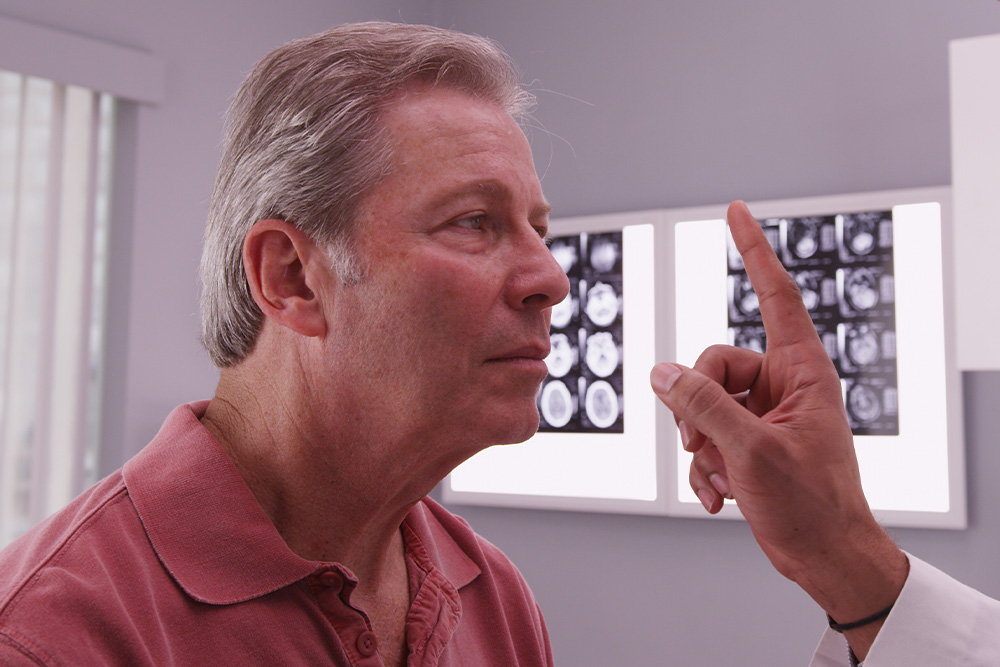
When most people think about vision problems, they picture issues with the eyes themselves. However, many visual difficulties actually begin in the brain. After a concussion, stroke, or other brain injury, the connection between the eyes and brain can be disrupted - causing symptoms that affect balance, reading, focus, and even daily comfort.
Brain Injuries and Vision
Your visual system is complex - about 70% of the brain is involved in vision processing. After a traumatic brain injury (TBI), concussion, or neurological event such as a stroke, the pathways that coordinate eye movement, focus, and perception can become impaired.
This can result in symptoms such as:
Blurred or double vision
Eye strain and headaches
Difficulty reading or focusing
Dizziness or balance problems
Light sensitivity
Problems with depth perception
These visual issues often persist long after other symptoms of a brain injury have improved, making specialized vision care essential for recovery.
What Is Neuro-Optometric Rehabilitation?
Neuro-optometric rehabilitation is a specialized form of vision therapy designed to retrain how the brain and eyes work together. Unlike traditional eye care that focuses on eyesight alone, this therapy targets the neurological processes behind vision. Our doctor evaluates how the eyes and brain communicate and creates a personalized treatment plan to rebuild these visual pathways. The goal is to restore comfort, coordination, and clarity - improving overall visual function and quality of life.

Dry eyes are one of the most common conditions that can affect our eyes and is estimated to affect millions of Americans. As you’ve probably guessed, dry eyes occur when tears fail to provide enough natural lubrication for the eyes to be comfortable and healthy. Exactly what causes dry eyes can vary significantly, from side effects from medications to prolonged computer use. What is clear is that while the condition isn’t sight-threatening, it can make day to day life much harder than it needs to be. Fortunately, there are treatments that can help, and arguably one of the most effective is Lipiflow.
What is Lipiflow?
Lipiflow is a new technological solution that addresses the underlying cause of your dry eyes, rather than simply treating the symptoms. It is most effective at helping patients whose dry eyes are caused by meibomian gland dysfunction – a condition characterized by problems with the way that the meibomian glands produce the oil that forms an essential part of our tear film. The meibomian glands can become less productive, or in some cases, even blocked by hardened oil deposits. This prevents the oil from reaching your tear film, making it less effective. Lipiflow targets the meibomian glands, warming them to break down oily blockages and massaging your eyes to make sure that the oil, and then the tear film, is evenly dispersed.
This helps to combat the symptoms associated with dry eyes, which can include:
Eye fatigue
Dry, scratchy and uncomfortable eyes
Blurred vision
Sensitivity to light
Difficulty wearing contact lenses
Your eye doctor will be able to advise you if Lipiflow has the potential to be a suitable solution for your dry eyes.

In the realm of eye care, surgical co-management has emerged as a collaborative approach that aims to provide patients with comprehensive and seamless treatment. This concept involves the joint efforts of optometrists and ophthalmologists, each bringing their unique expertise to the table. By working together, these eye care professionals strive to enhance patient outcomes and deliver exceptional care.
The Role of Optometrist and Ophthalmologist in Surgical Co-Management
Surgical co-management is built upon the unique skill sets and areas of expertise of optometrists and ophthalmologists. By understanding their respective roles, you can appreciate the synergy that this collaborative approach fosters.
Optometrists are primary eye care professionals who specialize in the examination, diagnosis, and non-surgical treatment of vision disorders. Their responsibilities in surgical co-management include:
Performing comprehensive eye examinations and evaluations
Monitoring and managing pre-existing eye conditions
Providing pre-operative and post-operative care
Educating patients on surgical procedures and aftercare
Collaborating with ophthalmologists to ensure continuity of care
Ophthalmologists are medical doctors who specialize in the diagnosis, treatment, and surgical management of eye diseases and disorders. Their role in surgical co-management encompasses:
Evaluating patients' candidacy for surgical interventions
Performing complex surgical procedures
Providing specialized medical and surgical care
Collaborating with optometrists to ensure seamless patient care
Monitoring and managing post-operative complications
By combining the expertise of optometrists and ophthalmologists, surgical co-management ensures that patients receive comprehensive and coordinated care throughout their treatment journey.
How Surgical Co-Management Works
Surgical co-management is a well-orchestrated process that involves several key steps. Understanding how it works can help you navigate this collaborative approach with confidence.
Initial Evaluation: The process typically begins with an optometrist conducting a comprehensive eye examination. During this evaluation, the optometrist assesses the patient's visual needs, identifies any potential issues, and determines if a surgical intervention is necessary.
Referral and Consultation: If surgery is recommended, the optometrist refers the patient to an ophthalmologist for further evaluation and consultation. This step ensures that the patient receives specialized medical advice and a thorough assessment of their suitability for the proposed surgical procedure.
Pre-operative Care: The optometrist plays a crucial role in providing pre-operative care, which may include managing any existing eye conditions, ensuring the patient understands the surgical process, and addressing any concerns or questions they may have.
Surgical Procedure: The ophthalmologist performs the necessary surgical intervention, leveraging their specialized training and expertise in surgical techniques.
Post-operative Care: After the surgery, the patient's care transitions back to the optometrist, who closely monitors the recovery process and provides post-operative care and management. This may involve follow-up appointments, monitoring for any complications, and ensuring the patient adheres to the prescribed treatment plan.
Ongoing Collaboration: Throughout the entire process, the optometrist and ophthalmologist maintain open communication and collaborate closely. This ensures that the patient's care is seamless, and any concerns or issues are promptly addressed by the appropriate healthcare professional.

Strabismus is a relatively common condition, affecting about 2-4% of the population worldwide. It is one of the most frequent eye disorders seen in children, though it can also develop in adults due to conditions like stroke or trauma. In the United States alone, it is estimated that around 12 million people are affected by strabismus, with varying degrees of severity. Early diagnosis and treatment are essential, as untreated strabismus in children can significantly impair visual development.
What is Strabismus?
Strabismus, commonly referred to as crossed eyes, is a condition in which the eyes do not properly align with each other when looking at an object. One eye may turn in, out, up, or down, while the other eye focuses correctly. This misalignment can be constant or intermittent and may affect one or both eyes. Strabismus occurs when the muscles controlling eye movement do not work together correctly, leading to difficulty in focusing both eyes on a single point. This condition can lead to visual issues such as double vision and, in some cases, can result in amblyopia (lazy eye) if left untreated.
How Does Strabismus Develop?
Strabismus develops when there is a disruption in the coordination of the muscles that control eye movement, leading to a misalignment of the eyes. This disruption can result from various causes, including genetic factors, neuromuscular conditions, or problems with the nerves that transmit signals to the eye muscles. In some cases, it may be present at birth, while in others, it can develop later in childhood or adulthood due to conditions such as cerebral palsy, Down syndrome, or even trauma to the eye or brain.
Signs and Symptoms of Strabismus
The primary symptom of strabismus is the visible misalignment of the eyes. However, there are other signs and symptoms that may indicate the presence of this condition:
Blurred or double vision
Headaches or eye strain
Difficulty with depth perception and spatial awareness
Reduced visual acuity in one eye
Frequent eye rubbing or squinting
Tilting or turning the head to compensate for the misalignment

Dry eye is a common condition that occurs when the eyes do not produce enough tears or when the tears evaporate too quickly. There are several factors that can contribute to dry eye, including environmental factors, hormonal changes, and certain medications. Additionally, conditions such as meibomian gland dysfunction and blepharitis can also lead to dry eye.
The Role of Meibomian Gland Dysfunction and Blepharitis in Dry Eye
Meibomian gland dysfunction occurs when the meibomian glands, which are responsible for producing the oily layer of tears, become blocked or do not function properly. This can result in the tears evaporating too quickly and not providing enough lubrication for the eyes. Blepharitis is an inflammation of the eyelids that can cause redness, itching, and a gritty sensation in the eyes. Both meibomian gland dysfunction and blepharitis can contribute to dry eye by disrupting the natural tear film.
Recognizing the Symptoms of Dry Eye
Dry eye can cause a variety of symptoms, which can vary in severity from person to person. Some common symptoms of dry eye include:
Dryness: The most common symptom of dry eye is a feeling of dryness or grittiness in the eyes. This can make it uncomfortable to wear contact lenses or spend long periods of time looking at a screen.
Redness: Dry eye can cause the blood vessels in the eyes to become more prominent, leading to redness and irritation.
Itching: Some people with dry eye may experience itching or a burning sensation in their eyes.
Excessive tearing: Dry eye can also cause excessive tearing. This is the body's response to the irritation caused by the lack of lubrication in the eyes.
If you are experiencing any of these symptoms, it is important to consult with an optometrist for a proper diagnosis and treatment plan.


Contact Info
Office Hours
- Monday Closed
- Tuesday 10:00am - 6:00pm
- Wednesday 10:00am - 6:00pm
- Thursday 10:00am - 7:00pm
- Friday 10:00am - 6:00pm
- Saturday 10:00am - 2:00pm
- Sunday Closed
© 2026 Hillsdale Eyecare. All Rights Reserved. Accessibility Statement | Privacy Policy | Sitemap
Powered by:
